

A comprehensive guide to adult otoplasty under local anaesthetic
Dr. Shailesh Vadodaria, Consultant Plastic Surgeon
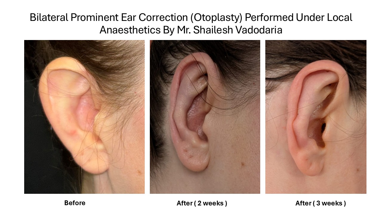
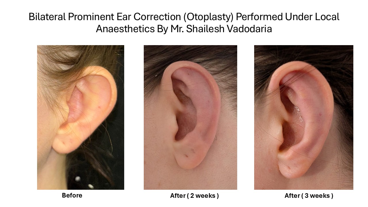
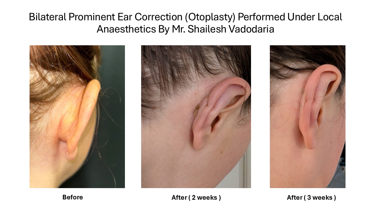
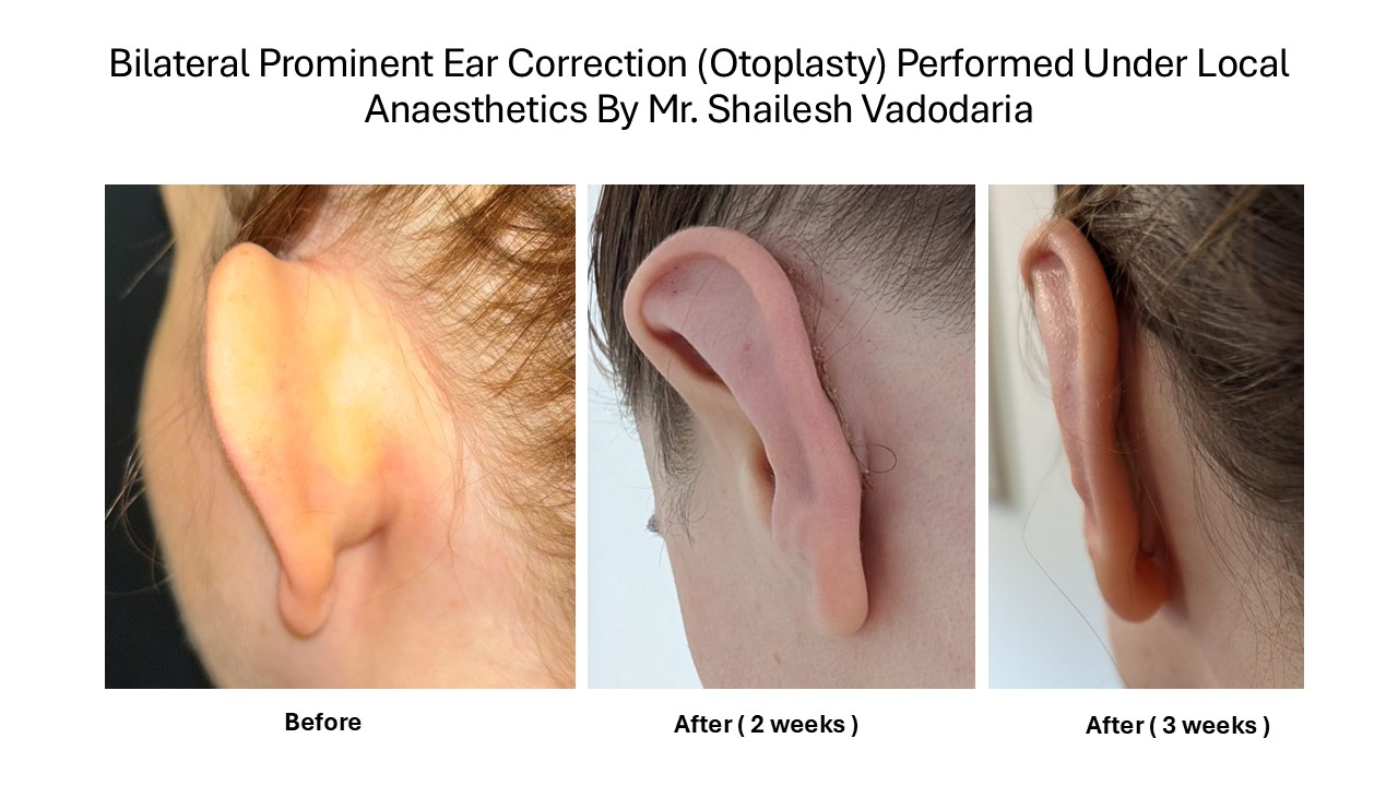
Prominent ears are a normal variation in appearance. Treatment is a personal choice. If their position bothers you, otoplasty can bring the ears closer to the head and improve their contours.
This guide explains ear anatomy, consultation, my preferred suture technique, local anaesthesia, recovery, possible complications and when to seek help. It is intended for adults considering treatment at MACS Clinic. It should help you prepare for an informed discussion; your examination and written care plan determine the treatment appropriate for you.
Your care at a glance
Initial enquiry and information pack, followed by a complimentary video consultation and photographs.
Face to face examination, medical assessment, personalised surgical plan and consent before treatment.
Usually about two hours for both ears, under local anaesthetic without sedation, as a day case.
Protective bandage for 48 hours, unless the surgeon advises otherwise; a soft headband at night for three months.
No swimming or contact sports for at least six weeks. Returning to contact sports needs individual clearance and may require longer.
Seek urgent assessment for severe or increasing pain, rapid swelling, spreading redness, discharge or fever.
General patient guidance draws on ISAPS and The Aesthetic Society, formerly the American Society for Aesthetic Plastic Surgery. Clinic timings and preferences describe the MACS pathway rather than a universal society protocol.
Understanding the external ear
Labelled external ear anatomy with arrows to the helix, antihelix, scapha, concha, tragus, antitragus, triangular fossa, lobule and canal entrance
The visible ear is the auricle, or pinna. Skin covers a flexible cartilage framework, except in the soft earlobe. Its folds and hollows create the familiar ear shape. The diagram is schematic: normal ears vary considerably.
Structure | Meaning |
Helical rim or helix | The curved outer edge of the ear. |
Antihelical fold or antihelix | The inner raised fold, which divides into two upper branches. Often called the antihelical rim. |
Scapha | The narrow hollow between the helix and antihelix. |
Concha | The bowl shaped hollow leading towards the ear canal. |
Tragus | The small projection immediately in front of the canal opening. |
Antitragus | The smaller projection opposite the tragus, just above the earlobe. |
Triangular fossa | The hollow between the two upper branches of the antihelix. |
Lobule | The soft earlobe, which has no cartilage framework. |
External auditory meatus | The entrance to the ear canal. |
Otoplasty chiefly changes the cartilage folds and the relationship between the concha and the side of the head. It is not an operation on the eardrum or middle ear and is not intended to improve hearing.