
AT A GLANCE
Milia (singular: milium) are tiny, benign cysts filled with keratin—the structural protein found in the outer skin. They are not infectious, are not “milk” or trapped sebum, and are usually an aesthetic concern rather than a health risk.
What are milia?
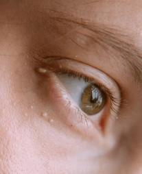
Milia are small, superficial keratin cysts lined by stratified squamous epithelium. They commonly measure about 1–3 mm and appear as firm, smooth, white, ivory or pearly dome-shaped bumps. They are usually painless and non-inflamed. The plural is “milia”; one lesion is a “milium”.
Although often called “milk spots”, milia do not contain milk. They are also not the same as acne whiteheads: a typical milium is a closed cyst without an open pore, so forceful squeezing commonly fails and may injure the skin.
Where do they commonly form?
In adults and older children, primary milia favour facial skin bearing fine (vellus) hairs—particularly the lower eyelids, upper cheeks, temples and forehead. They may also occur around the nose, on the scalp, behind the ears and, less commonly, on the trunk or genital skin. Newborn milia commonly affect the nose and central face and usually settle spontaneously.
Why do milia form?
Primary (de novo) milia
Primary milia arise without an obvious preceding injury. Keratin becomes enclosed within a tiny superficial epithelial cyst. Histological studies suggest that many primary lesions relate to the lower portion of the follicular infundibulum/sebaceous collar of fine hair follicles. Exactly why an individual lesion forms is not always identifiable.
Secondary milia after trauma, inflammation or surgery
Secondary milia form while skin is healing or remodelling after damage to the epidermis or skin appendages. Reported settings include burns, blistering disorders, inflammatory rashes, abrasions, graft donor sites, dermabrasion, laser resurfacing, radiotherapy and surgical scars. They may appear weeks or months after the original event, sometimes in a line or cluster within the healed area. Experimental and histological work suggests that regenerating epidermis, eccrine ducts and/or damaged follicular structures can become enclosed and produce a keratin cyst.
Medication-associated secondary milia have also been reported, including with some topical corticosteroids and certain systemic medicines. An association in one person does not prove that a product caused the lesions, and prescribed medication should not be stopped without advice.
AFTER SURGERY A small white bump in or near a healed scar may be a secondary milium, but suture reaction, a tiny epidermoid cyst, infection and other lesions can look similar. Examination is advisable before treatment—particularly if the area is red, painful, draining or enlarging. |
Clinical appearance and diagnosis
- One or more discrete, smooth, firm, white-to-yellow pearly papules, usually 1–3 mm.
- Usually no pain, itch, redness or discharge, although uncommon eruptive variants can itch.
- Often clustered around the eyelids and cheeks; secondary lesions follow the distribution of an earlier injury or scar.
- Diagnosis is usually clinical. Dermoscopy or, rarely, biopsy may be used when the diagnosis is uncertain.
Conditions that can resemble milia include closed comedones, sebaceous hyperplasia, syringomas, molluscum contagiosum, xanthelasma, calcinosis cutis and small epidermoid cysts. “Colloid milium” is a different sun-related deposition disorder despite the similar name.
Feature | Typical milium | Closed comedone |
Contents | Compact keratin pearl | Keratin/sebum within a blocked follicle |
Surface opening | Usually none | Follicular opening is obstructed |
Feel/look | Firm, pearly, discrete | Skin-coloured or white acne bump |
Best approach | Confirm diagnosis; observe or clinician extraction | Acne skincare/medical treatment according to severity |
This comparison is a guide only; clinical examination is sometimes needed because several benign lesions can appear similar
Do milia always need treatment?
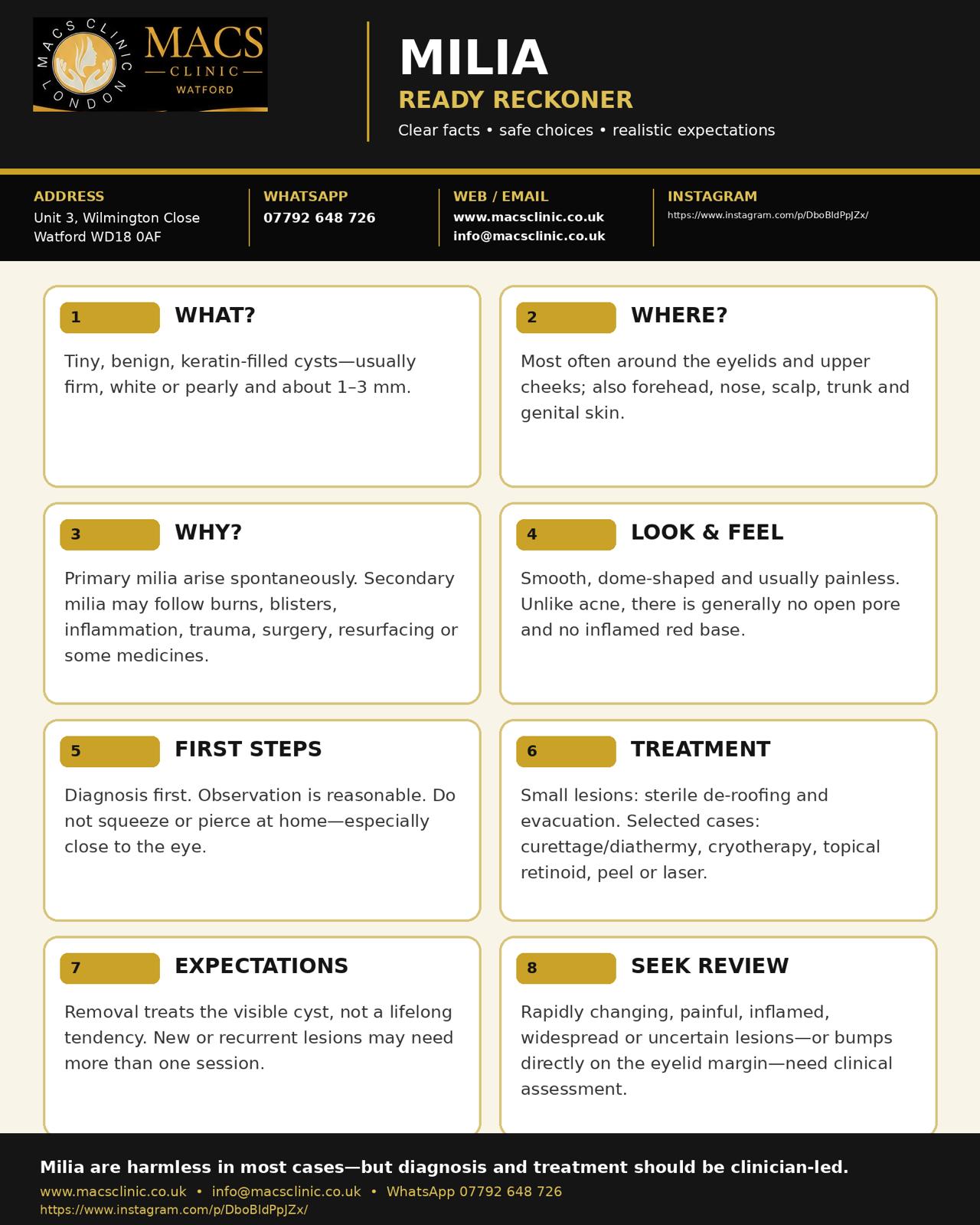
No. Milia are benign and treatment is optional. Neonatal milia almost always clear without intervention. Some adult primary lesions resolve, but adult and secondary milia may persist. Treatment is reasonable when the diagnosis is secure and the lesions are cosmetically troublesome, recurrently irritated or of particular concern to the patient.
Evidence-based treatment options
1. Sterile de-roofing and evacuation
For a small number of accessible lesions, a trained clinician can cleanse the skin, create a minute opening in the cyst roof with a sterile needle or fine blade, and gently express or lift out the keratin pearl. This is the most direct and commonly described treatment. Delicate technique is especially important around the eyelids.
Local anaesthetic is not always needed for a tiny superficial lesion but may be considered according to site, number, patient comfort and clinician preference. The procedure should not be attempted at home.
2. Curettage, electrosurgery/diathermy or cryotherapy
Selected lesions can be treated using curettage, low-energy electrosurgery/diathermy or cryotherapy. These methods can be useful when simple evacuation is unsuitable, but they create a controlled skin injury and may carry a greater risk of pigment change or scarring—particularly on eyelid skin and in darker skin tones.
3. Topical retinoids
A clinician may prescribe a topical retinoid, such as tretinoin, for numerous or eruptive milia. Retinoids normalise keratinisation and may reduce lesions or make extraction easier. Evidence for milia is mainly from case reports and small series, not large comparative trials. Irritation, dryness and photosensitivity are common; retinoids must be used carefully near the eyes and require pregnancy-related precautions.
4. Chemical peels and dermabrasion
Superficial chemical peeling or dermabrasion has been reported for extensive or resistant milia, usually as a specialist treatment. These approaches are not first-line for a few isolated lesions. Risks include irritation, prolonged redness, infection, post-inflammatory hyperpigmentation or hypopigmentation, and scarring.
5. Laser treatment
Carbon dioxide and erbium:YAG laser ablation have been reported for extensive milia, multiple eruptive milia and milia en plaque. Published evidence is largely limited to case reports and small case series, so potential benefit must be balanced against cost, downtime, pigment alteration, burns and scarring. Eye protection and appropriately trained treatment are essential near the orbit.
6. Treatment of an underlying condition
When milia follow a blistering or inflammatory skin disorder, reducing ongoing skin injury and treating the underlying condition may help prevent further lesions. Rare variants such as milia en plaque or widespread eruptive milia merit dermatology assessment. Antibiotics such as minocycline have been reported for milia en plaque, but this is not routine treatment for ordinary isolated milia and the evidence is limited.
What to expect after de-roofing
- A pinpoint opening, mild redness, tenderness or a tiny crust for several days.
- Keep the area clean and follow the clinician’s aftercare instructions; avoid picking.
- Use suitable sun protection after healing, particularly if prone to pigmentation.
- Contact the clinic if redness, swelling, pain, heat, discharge or visual symptoms worsen.
Possible complications
Complications are uncommon when treatment is appropriate and carefully performed, but can include pain, bleeding, bruising, infection, delayed healing, persistent redness, post-inflammatory hyperpigmentation or hypopigmentation, a small scar, incomplete removal and recurrence. Procedures close to the eye carry additional risks and require particular anatomical expertise. Treating a lesion that has been incorrectly diagnosed may delay the correct diagnosis.
Can milia recur?
Yes. A successfully evacuated cyst is removed, but another milium can form nearby or a residual cyst wall/content can produce persistence. Patients who develop multiple lesions, or whose milia follow ongoing irritation or skin disease, may require staged or repeat treatment. Recurrence does not usually imply anything dangerous.
Can they be prevented?
There is no guaranteed prevention. Sensible measures include avoiding picking or unnecessary skin trauma, using prescribed topical steroids only as directed, choosing non-irritating skincare, and using sun protection. Evidence that any particular cosmetic product reliably prevents milia is limited. Heavy products may be associated with milia in some people, but this is not universal.
When should you seek medical advice?
- The diagnosis is uncertain or the lesion is changing, pigmented, ulcerated or bleeding.
- The bump is painful, red, hot, draining or rapidly enlarging.
- There are many sudden-onset lesions, significant itching, blistering or another rash.
- The lesion sits on the eyelid margin or close to the eye.
- Milia repeatedly form within a surgical scar or after a skin procedure.
MACS CLINIC PERSPECTIVE For a small, typical milium, sterile de-roofing and gentle evacuation is often a simple and effective approach. The priority is confirming the diagnosis, choosing the least traumatic appropriate treatment, and setting realistic expectations about recurrence and the possibility of more than one session. |
Frequently asked questions
Are milia contagious?
No. They are not caused by an infection and cannot be passed to another person.
Are milia dangerous?
Ordinary milia are benign. Assessment matters when the appearance is atypical or the diagnosis is uncertain.
Can I squeeze one?
It is best not to. A milium has no easy surface opening; squeezing can cause bruising, inflammation, infection, pigment change or a scar.
Will skincare dissolve it?
Some superficial or widespread lesions may improve with clinician-guided retinoid treatment, but a mature isolated keratin cyst often needs direct evacuation if removal is desired.
How many sessions will I need?
One session may be sufficient for a few lesions, but numerous, secondary or recurrent milia may need staged or repeat treatment.
Important note
This blog provides general education and cannot diagnose an individual skin lesion or replace a clinical consultation. Treatment choice depends on diagnosis, site, skin type, medical history, medicines, previous procedures and personal priorities. Evidence for uncommon or extensive milia treatments is often limited to case reports or small series.
Contact MACS Clinic
- Phone: 020 7078 4378
- WhatsApp: 07792 648 726
- Email: enquiries@macsclinic.co.uk
- Website: www.macsclinic.co.uk
- BOOK a FREE Video Consultation: https://calendly.com/macsclinic/free-video-consultation?month=2025-01