Seborrhoeic keratosis is one of the most common benign (non-cancerous) skin growths seen in adults. It is sometimes referred to as a “senile wart” or “age spot”, although it has nothing to do with infection or poor hygiene.
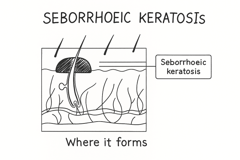
These lesions arise from the epidermis (the outer layer of the skin) and are made up of proliferating keratinocytes—the same cells that form healthy skin.
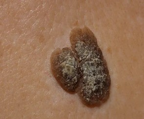
What Does Seborrhoeic Keratosis Look Like?
Typical features include:
- Colour: skin-coloured, light brown, dark brown, grey or black
- Surface: waxy, rough, “stuck-on” or warty appearance
- Shape: round or oval, sharply defined
- Consistency: can be crumbly or flaky
- Number: single or multiple lesions
They commonly appear on the:
- face
- neck
- chest
- back
- shoulders and upper limbs
They do not occur on the palms or soles.
Who Gets Seborrhoeic Keratosis?
- Age: Most common after 50 years, increases with age
- Gender: Affects both men and women equally
- Ethnicity: Seen in all skin types; can be more numerous in darker skin tones
- Genetics: Tends to run in families
- Other factors: Sun exposure may contribute, but SKs can occur on covered skin too
They are not contagious and not related to skin cancer, although they can sometimes resemble one.
Clinical Diagnosis
Diagnosis is usually made by visual examination. Key clinical criteria include:
- “stuck-on”, waxy appearance
- well-defined edge
- warty/rough surface
- dull, not inflamed (unless irritated)
- history of gradual growth
Differential Diagnosis
Conditions that can resemble seborrhoeic keratosis:
- Melanoma (most important to differentiate)
- Pigmented basal cell carcinoma
- Solar lentigo (sun spot)
- Common wart (viral wart)
- Dermatosis papulosa nigra (common in darker skin types)
Investigations
- Most cases do not require tests
- Biopsy / histopathology is performed when:
- the diagnosis is uncertain
- the lesion looks atypical
- there is rapid growth, bleeding or ulceration
- there is concern for melanoma or other skin cancer
- the diagnosis is uncertain
It is advisable to send tissue for pathology to confirm the diagnosis
Treatment Options
Seborrhoeic keratosis is benign, so treatment is usually for:
- cosmetic reasons
- irritation (e.g., friction from clothing)
- bleeding or inflammation
- diagnostic uncertainty
Treatment techniques include:
Method | Key Points |
Surgical shave excision | Allows histology; local anaesthetic; good cosmetic result |
Curettage | Scraping technique; often combined with cautery |
Cryotherapy (liquid nitrogen) | No specimen for pathology; may lighten/darken skin |
Electrocautery/laser | Destroys lesion; no tissue available for testing |
Surgical excision preferred if there is any doubt about diagnosis so tissue can be sent to pathology.
When To See a Doctor
Seek medical review if a lesion:
- changes in shape, size or colour
- becomes rapidly larger
- bleeds spontaneously
- appears very dark or irregular
- causes persistent irritation
Any suspicious lesion should be examined to exclude any skin cancer
Key Takeaways
- SK is a very common benign skin lesion
- Not cancerous, not contagious
- More common with age and family history
- Can resemble melanoma — always check if unsure
- Surgical removal allows histopathological confirmation
Excellent outcomes when treated properly
Contact MACS Clinic
- Phone: 020 7078 4378
- WhatsApp: 07792 648 726
- Email: enquiries@macsclinic.co.uk
- Website: www.macsclinic.co.uk
BOOK a FREE Video Consultation: https://calendly.com/macsclinic/free-video-consultation?month=2025-01