
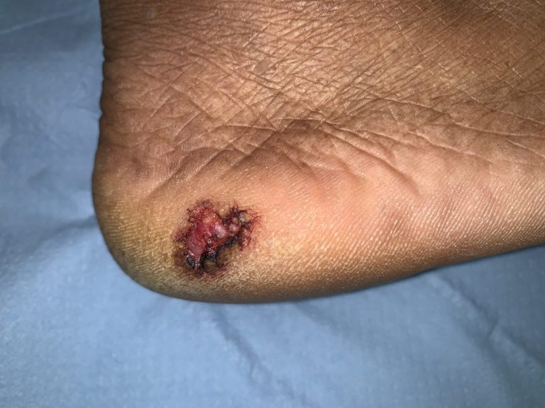
Non-healing ulcers, especially on the sole of the foot, are a serious medical concern due to their potential complications and the risk of underlying conditions that may impair healing. While many foot ulcers are benign and arise from common causes like diabetes, injury, pressure or circulatory problems, they can also signify something far more dangerous—Marjolin’s ulcer, a rare but aggressive form of skin cancer.
What is a Foot Ulcer?
A foot ulcer is an open sore or wound that develops on the skin of the foot, often caused by prolonged pressure, trauma, or poor circulation. Foot ulcers can vary in depth and severity, and they are particularly common in individuals with diabetes, vascular diseases, or reduced mobility. Ulcers may heal on their own with proper care, but when they don’t, it becomes crucial to identify the cause of the delayed healing.
Common Causes of Foot Ulcers Include:
- Diabetic Foot Ulcers: Caused by neuropathy (nerve damage) and reduced blood flow, common in diabetic patients.
- Pressure Ulcers: Occurring in areas of constant pressure, such as the heel or ball of the foot, especially in bedridden or wheelchair-bound individuals.
- Venous and Arterial Ulcers: Linked to poor circulation in the legs and feet due to vein or artery disease.
- Trauma: Repeated injury or constant pressure on a specific area of the foot can break the skin and lead to an ulcer.
While most foot ulcers can heal with proper treatment e.g. regular change of dressings, non-healing ulcers—those that persist for months or years—can indicate more severe underlying problems, including Marjolin’s ulcer/ invasive cancer.
What Is Marjolin’s Ulcer?
Marjolin’s ulcer is a malignant transformation that occurs in long-standing wounds, scars, or non-healing ulcers, turning into squamous cell carcinoma (SCC), a form of skin cancer. Though uncommon, Marjolin’s ulcer is highly aggressive, and its ability to invade surrounding tissue and spread to other parts of the body (metastasize) makes it critical to catch early.
In cases of chronic foot ulcers, especially those linked to previous burns, trauma, or scars, the risk of malignant transformation increases, making early detection and biopsy essential.
Warning Signs of Marjolin’s Ulcer
Identifying Marjolin’s ulcer early is key to successful treatment. Patients with long-standing ulcers should watch for the following signs:
- Rapid Growth: A sudden increase in the size of the ulcer.
- Appearance Changes: Alterations in colour, texture, or surface appearance.
- Unusual Discharge: Excessive bleeding, pus, or a foul odor.
- Pain: Pain that feels out of proportion to the ulcer’s appearance.
- New Growth: Nodules or growths within or around the ulcer site.
If any of these warning signs are present, immediate seek medical advice to rule out cancerous changes.
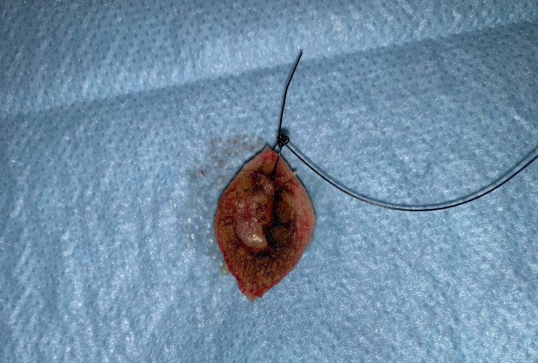
The Role of Biopsy in Diagnosing Marjolin’s Ulcer
The most reliable method of diagnosing Marjolin’s ulcer is a biopsy. A biopsy involves taking a small sample of the ulcer tissue, which is then examined under a microscope to look for cancerous cells.
Ruling Out Malignancy: A biopsy determines whether the ulcer is undergoing malignant changes. Early detection of squamous cell carcinoma can save lives.
If the biopsy confirms, Marjolin’s ulcer then the matter needs discussion in multidisciplinary team for necessity of further investigations, staging of disease and combined discussion for wide local removal of the cancer, reconstruction and if required chemotherapy/radiotherapy.
Preventing Delays: Prompt biopsy ensures that potential skin cancers are diagnosed early, leading to better outcomes and preventing life-threatening complications.
Suggested Approach For Foot Ulcer Treatment
The surgeon assess all contributing factors, including vascular insufficiency, infections, and pressure points, to decide a treatment plan.
The surgeon and his team use debridement, specialized dressings, and offloading techniques (to relieve pressure) to promote healing.
When a non-healing ulcer raises suspicion of malignant transformation, the surgeon would perform a biopsy.
If cancer is detected, the surgeon will refer the patient to skin cancer multidisciplinary team. If the non-healing ulcer is non-cancerous then several surgical techniques are available, depending on the size, depth, location of the ulcer, and underlying causes.
Surgical Treatments for Non-Healing Ulcers
- Debridement
Debridement involves the surgical removal of dead, damaged, or infected tissue from the ulcer. This helps promote healing by:
- Reducing the bacterial load.
- Removing necrotic tissue that impairs healing.
- Stimulating the formation of healthy tissue.
Debridement can be performed using several techniques, including sharp debridement (using surgical instruments like scalpels) and mechanical debridement (using hydrotherapy or wound irrigation). In some cases, enzymatic debridement with topical medications is also combined with surgical approaches.
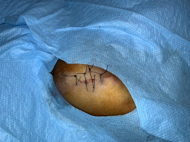
- Reconstruction
For large ulcers that cannot heal on their own, The skin graft involves covering the ulcer with healthy skin taken from another part of the body (the donor site). Grafting is useful for promoting the growth of new skin and protecting the underlying structures of the foot.
Types of skin grafts include:
- Split-thickness skin grafts: A thin layer of skin is taken from the donor site and applied to the ulcer.
- Full-thickness skin grafts: A thicker layer of skin is used, often for more complex ulcers requiring more coverage.
- Flap Surgery
When skin grafts are not sufficient, flap surgery can be an option. Flap surgery involves transferring a section of tissue (including skin, fat, muscle, and blood vessels) from one part of the body to the ulcerated area. This is particularly useful for deep ulcers or those with exposed bones or tendons.
Types of flap surgery include:
- Local flaps: Tissue from nearby the ulcer site is used to cover the area.
- Free flaps: Tissue from a distant part of the body is moved to the ulcer site, along with its blood supply.
For the treatment of long-standing, non-healing ulcers, you have a few options:
- Contact Your GP: If you are experiencing a non-healing ulcer, your first step should be to contact your general practitioner (GP) for an initial assessment and treatment options.
- Insured Patients: MACS Clinic provides services for patients insured with WPA, CIGNA, Vitality, BUPA, AVIVA and BUPA International. Consultation (video and/or face to face) for non-insured patient is complimentary (free). Your GP can provide a referral, and MACS Clinic will handle your treatment which will be covered by your insurance plan.
- Private Patients: If you are opting for private treatment, after your complimentary/free consultation at MACS Clinic, Mr. Vadodaria will provide you with a treatment plan and a detailed quote for your treatment.
What post-surgical care needs to be taken after the Ulcer removal procedure at MACS Clinic?
- Regular check-ups to ensure the wound is healing as expected. A wound check with the surgeon or his team is scheduled within 7-10 days, followed by a follow-up visit after six weeks to ensure proper recovery.
- Use specialized shoes, braces, or cushions to relieve pressure from the ulcer site.
- Antibiotics and painkillers are prescribed for 5 to 7 days to prevent infection and manage any discomfort.
Contact Us
call us
02070784378
Email Us
info@macsclinic.co.uk
Opening Hours
9:30 AM - 18:00 PM
Visit Us
Unit 3, Wilmington Close Watford WD18 0AF